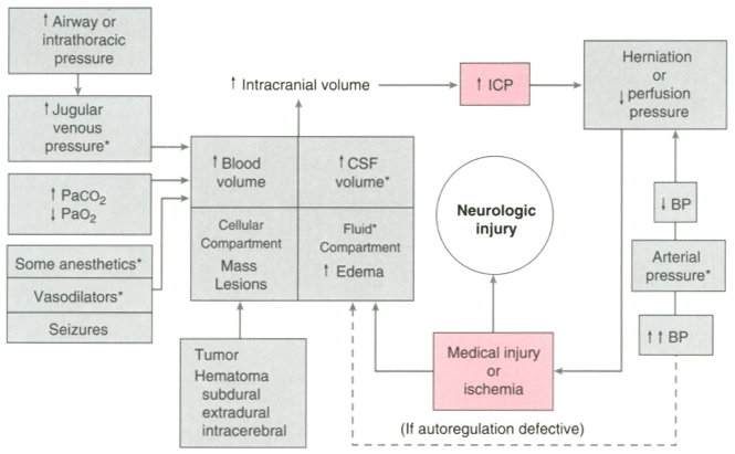
Figure 53-4
Pathophysiology of intracranial hypertension. The figure
depicts the manner in which increases in the volumes of any or all of the four intracranial
compartments, blood, cerebrospinal fluid (CSF), fluid (interstitial or intracellular),
and cells (four-part rectangle) result in increases in intracranial pressure and
eventual neurologic damage. Elements that are potentially under control of the anesthesiologist
are indicated by asterisks. (Control of CSF volume
requires the presence of a ventriculostomy catheter.) The herniation pathways are
depicted in Figure 53-1
.