Abnormal Central Venous Pressure Waveforms
Various pathophysiologic conditions may be diagnosed or confirmed
by examination of the CVP waveform ( Table
32-9
). One of the most common applications is the rapid diagnosis of cardiac
arrhythmias.[311]
In atrial
fibrillation ( Fig. 32-25
),
the a wave disappears and the c wave becomes more prominent because atrial volume
is greater at end-diastole and the onset of systole owing to the absence of effective
atrial contraction. Occasionally, atrial fibrillation or flutter waves may be seen
in the CVP trace,
TABLE 32-9 -- Central venous pressure waveform abnormalities
|
Condition |
Characteristics |
|
Atrial fibrillation |
Loss of a wave |
|
Prominent c wave |
|
Atrioventricular dissociation |
Cannon a wave |
|
Tricuspid regurgitation |
Tall systolic c-v wave |
|
Loss of x descent |
|
Tricuspid stenosis |
Tall a wave |
|
Attenuation of y descent |
|
Right ventricular ischemia |
Tall a and v waves |
|
Steep x and y descents |
|
M or W configuration |
|
Pericardial constriction |
Tall a and v waves |
|
Steep x and y descents |
|
M or W configuration |
|
Cardiac tamponade |
Dominant x descent |
|
Attenuated y descent |
|
Respiratory variation during spontaneous or positive-pressure
ventilation |
Measure pressures at end-expiration |
when the ventricular rate is slow. Isorhythmic atrioventricular
dissociation or junctional (nodal) rhythm
(see Fig. 32-25
) alters
the normal sequence of atrial contraction before ventricular contraction. Instead,
atrial contraction now occurs during ventricular systole, when the tricuspid valve
is closed, thereby inscribing a tall cannon a wave in the CVP waveform. Absence
of normal atrioventricular synchrony during ventricular pacing (see Fig.
32-25
) can be identified in a similar fashion by searching for cannon waves
in the venous pressure trace. In these instances, CVP helps diagnosis the cause
of arterial hypotension; loss of the normal end-diastolic atrial kick may not be
as evident in the ECG trace as it is in the CVP waveform.
Right-sided valvular heart diseases alter the CVP waveform in
different ways.[312]
Tricuspid
regurgitation ( Fig. 32-26
)
produces abnormal systolic filling of the right atrium through the incompetent valve.
A broad, tall systolic c-v wave is inscribed that begins in early systole and obliterates
the systolic x descent in atrial pressure. The CVP trace is said to be ventricularized
because it resembles right ventricular pressure. Note that this regurgitant wave
differs in onset, duration, and magnitude from a normal CVP v wave caused by end-systolic
atrial filling from the venae cavae. In patients with tricuspid regurgitation, right
ventricular end-diastolic pressure is overestimated by the numeric display on the
bedside monitor, which reports a single mean value for CVP. Instead, right ventricular
end-diastolic pressure is estimated best by measuring the CVP value at the time of
the ECG R wave, before the regurgitant systolic wave (see Fig.
32-26
). Unlike tricuspid regurgitation, tricuspid
stenosis (see Fig. 32-26
)
is a diastolic defect in atrial emptying and ventricular filling. Mean CVP is elevated,
and a pressure gradient exists throughout diastole between the right atrium and ventricle.
The a wave is unusually prominent and the y descent is attenuated because of the
impaired diastolic egress of blood from the atrium. Other conditions that
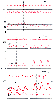 Figure 32-25
Central venous pressure (CVP) changes caused by cardiac
arrhythmias. A, Atrial fibrillation. Note the absence
of the a wave, a prominent c wave, and a preserved v wave and y descent. This arrhythmia
also causes variation in the electrocardiographic (ECG) R-R interval and left ventricular
stroke volume, which can be seen in the ECG and arterial pressure (ART) traces.
B, Isorhythmic atrioventricular dissociation. In
contrast to the normal end-diastolic a wave in the CVP trace (left
panel), an early systolic cannon wave is inscribed (asterisk,
right panel). The reduced ventricular filling accompanying this arrhythmia
causes decreased arterial blood pressure. C, Ventricular
pacing. Systolic cannon waves are evident in the CVP trace during ventricular pacing
(left panel). Atrioventricular sequential pacing
restores the normal venous waveform and increases arterial blood pressure (right
panel). The ART scale is shown on the left,
the CVP scale on the right. (Redrawn from
Mark JB: Atlas of Cardiovascular Monitoring. New York, Churchill Livingstone, 1998,
Figs. 14-1, 14-5, and 14-16.)
Figure 32-25
Central venous pressure (CVP) changes caused by cardiac
arrhythmias. A, Atrial fibrillation. Note the absence
of the a wave, a prominent c wave, and a preserved v wave and y descent. This arrhythmia
also causes variation in the electrocardiographic (ECG) R-R interval and left ventricular
stroke volume, which can be seen in the ECG and arterial pressure (ART) traces.
B, Isorhythmic atrioventricular dissociation. In
contrast to the normal end-diastolic a wave in the CVP trace (left
panel), an early systolic cannon wave is inscribed (asterisk,
right panel). The reduced ventricular filling accompanying this arrhythmia
causes decreased arterial blood pressure. C, Ventricular
pacing. Systolic cannon waves are evident in the CVP trace during ventricular pacing
(left panel). Atrioventricular sequential pacing
restores the normal venous waveform and increases arterial blood pressure (right
panel). The ART scale is shown on the left,
the CVP scale on the right. (Redrawn from
Mark JB: Atlas of Cardiovascular Monitoring. New York, Churchill Livingstone, 1998,
Figs. 14-1, 14-5, and 14-16.)
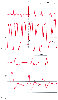 Figure 32-26
Central venous pressure (CVP) changes in tricuspid valve
disease. A, Tricuspid regurgitation increases mean
CVP, and the waveform displays a tall systolic c-v wave that obliterates the x descent.
In this example, the a wave is not seen because of atrial fibrillation. Right ventricular
end-diastolic pressure is estimated best at the time of the electrocardiographic
R wave (arrows) and is lower than mean CVP. B,
Tricuspid stenosis increases mean CVP, the diastolic y descent is attenuated, and
the end-diastolic a wave is prominent. (Redrawn from Mark JB: Atlas of
Cardiovascular Monitoring. New York, Churchill Livingstone, 1998, Figs. 17-3 and
17-15.)
Figure 32-26
Central venous pressure (CVP) changes in tricuspid valve
disease. A, Tricuspid regurgitation increases mean
CVP, and the waveform displays a tall systolic c-v wave that obliterates the x descent.
In this example, the a wave is not seen because of atrial fibrillation. Right ventricular
end-diastolic pressure is estimated best at the time of the electrocardiographic
R wave (arrows) and is lower than mean CVP. B,
Tricuspid stenosis increases mean CVP, the diastolic y descent is attenuated, and
the end-diastolic a wave is prominent. (Redrawn from Mark JB: Atlas of
Cardiovascular Monitoring. New York, Churchill Livingstone, 1998, Figs. 17-3 and
17-15.)
reduce right ventricular compliance, such as right ventricular ischemia, pulmonary
hypertension, or pulmonic valve stenosis, may produce a prominent end-diastolic a
wave in the CVP trace but do not attenuate the early diastolic y descent. CVP waveform
morphology changes in other characteristic ways in the presence of pericardial diseases
and right ventricular infarction. These patterns are interpreted best in conjunction
with PAP monitoring, which is discussed in the next section.
Perhaps the most important application of CVP monitoring is to
provide an estimate of the adequacy of circulating blood volume and right ventricular
preload. As noted earlier, for this purpose, transmural CVP is always the pressure
of physiologic interest. In clinical practice, however, we measure and record pressures
referenced to ambient atmospheric pressure. Consequently, accurate interpretation
of CVP requires the physician to consider alterations in intrathoracic or juxtacardiac
pressure that occur during the respiratory cycle.[294]
[303]
During spontaneous breathing ( Fig.
32-27
), inspiration causes a decrease in pleural and juxtacardiac pressure
that is transmitted, in part, to the right atrium and lowers CVP. This same decrease
in pleural pressure will influence other measured central vascular pressures in similar
fashion. Note a subtle, but critically important observation about the measurement
of central vascular pressures. Although CVP measured relative to atmospheric pressure
decreases during the inspiratory phase of spontaneous ventilation, transmural CVP,
the difference between right atrial pressure and juxtacardiac pressure, may actually
increase slightly as more blood is drawn into the right atrium. The opposite pattern
is observed during positive-pressure ventilation, in which inspiration increases
intrathoracic pressure, raises the measured CVP, but decreases transmural CVP because
the elevated intrathoracic pressure reduces venous return. In clinical practice,
transmural pressures are rarely measured because of difficulty assessing juxtacardiac
or intrathoracic pressure.
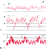 Figure 32-27
Respiratory influences on the measurement of central
venous pressure (CVP). A, During spontaneous ventilation,
the onset of inspiration (arrows) causes a reduction
in intrathoracic pressure that is transmitted to both the CVP and the pulmonary artery
pressure (PAP) waveforms. CVP should be recorded at end-expiration (mean CVP, 14
mm Hg). B, During positive-pressure ventilation,
the onset of inspiration (arrows) causes an increase
in intrathoracic pressure. CVP is still recorded at end-expiration (mean CVP, 8
mm Hg). (Redrawn from Mark JB: Atlas of Cardiovascular Monitoring. New
York, Churchill Livingstone, 1998, Figs. 16-1 and 16-2.)
Figure 32-27
Respiratory influences on the measurement of central
venous pressure (CVP). A, During spontaneous ventilation,
the onset of inspiration (arrows) causes a reduction
in intrathoracic pressure that is transmitted to both the CVP and the pulmonary artery
pressure (PAP) waveforms. CVP should be recorded at end-expiration (mean CVP, 14
mm Hg). B, During positive-pressure ventilation,
the onset of inspiration (arrows) causes an increase
in intrathoracic pressure. CVP is still recorded at end-expiration (mean CVP, 8
mm Hg). (Redrawn from Mark JB: Atlas of Cardiovascular Monitoring. New
York, Churchill Livingstone, 1998, Figs. 16-1 and 16-2.)
Instead, end-expiratory values for cardiac filling pressure should be recorded in
all patients to provide the best estimate of transmural pressure. At the end of
expiration, intrathoracic and juxtacardiac pressures approach atmospheric pressure,
whether the patient is breathing spontaneously or receiving positive-pressure mechanical
ventilation (see Fig. 32-27
).
Proper pressure values can be determined by visual inspection of the CVP waveform
on a calibrated monitor screen or paper recording. Under most circumstances, transmural
CVP and the end-expiratory value for CVP will be close to one another. This facilitates
comparison of CVP values (and other cardiac filling pressures) obtained from the
same patient under varying patterns of ventilation, a common situation in anesthesia
and critical care.
Not only can individual CVP waveforms provide unique diagnostic
clues about the circulation, but trends in CVP during anesthesia and surgery are
also useful in estimating fluid or blood loss and guiding replacement therapy. It
is important to remember that the range in normal values is considerable and that
small changes in CVP may reflect significant changes in circulating blood volume
and right ventricular preload. Additional useful information may be derived from
examining how a fluid bolus simultaneously alters CVP and other variables of clinical
interest such as blood pressure, urine output, and so forth.