Normal Central Venous Pressure Waveforms
Strictly speaking, CVP is the pressure at the junction of the
venae cavae and right atrium and reflects the driving force for filling the right
atrium and ventricle. Because the large
TABLE 32-7 -- Normal cardiovascular pressures
|
Pressure |
Average (mm Hg) |
Range (mm Hg) |
|
Right Atrium |
|
|
|
a wave |
6 |
2–7 |
|
v wave |
5 |
2–7 |
|
Mean |
3 |
1–5 |
|
Right Ventricle |
|
|
|
Peak systolic |
25 |
15–30 |
|
End-diastolic |
6 |
1–7 |
|
Pulmonary Artery |
|
|
|
Peak systolic |
25 |
15–30 |
|
End-diastolic |
9 |
4–12 |
|
Mean |
15 |
9–19 |
|
Pulmonary Artery Wedge |
|
|
|
Mean |
9 |
4–12 |
|
Left Atrium |
|
|
|
a wave |
10 |
4–16 |
|
v wave |
12 |
6–21 |
|
Mean |
8 |
2–12 |
|
Left Ventricle |
|
|
|
Peak systolic |
130 |
90–140 |
|
End-diastolic |
8 |
5–12 |
|
Central Aorta |
|
|
|
Peak systolic |
130 |
90–140 |
|
End-diastolic |
70 |
60–90 |
|
Mean |
90 |
70–105 |
veins of the thorax, abdomen, and proximal ends of the extremities form a compliant
reservoir for a sizable percentage of the total blood volume, CVP is highly dependent
on the intravascular blood volume and intrinsic vascular tone of these capacitance
vessels. In other words, CVP or right atrial pressure reflects the appropriateness
of the blood volume to the capacity of the venous system.[304]
In addition to providing a measure of the circulating blood volume, CVP reflects
the functional capacity of the right ventricle. Based on the Frank-Starling mechanism,
higher right heart filling pressures are required to maintain ventricular stroke
output when right ventricular contractility is impaired. Thus, in clinical practice,
CVP monitoring is used for assessment of blood volume and right heart function.[305]
Normal CVP in an awake, spontaneously breathing patient ranges between 1 and 7 mm
Hg.
Normal mechanical events of the cardiac cycle are responsible
for the sequence of waves seen in a typical CVP trace. The CVP waveform consists
of five phasic events, three peaks (a, c, v) and two descents (x, y) ( Table
32-8
, Fig. 32-24
).
[306]
[307]
[308]
The most prominent wave is the a wave of atrial
contraction, which occurs at end-diastole after the ECG P wave. The a wave increases
atrial pressure and provides the "atrial kick" to fill the right ventricle through
the open tricuspid valve. Atrial pressure decreases after the a wave as the atrium
relaxes. This smooth decline in pressure is interrupted by the c
wave. This wave is a transient increase in atrial pressure produced by
isovolumic ventricular contraction, which closes the tricuspid valve and displaces
it toward the atrium. The c wave always follows the ECG R wave because it is generated
during the onset of ventricular systole. (Note that the c wave observed in a jugular
venous pressure trace might have a slightly more complex origin. This wave has been
attributed to early systolic pressure transmission from the adjacent carotid artery
and may be termed a carotid impact wave.[309]
Because
jugular venous pressure also reflects right atrial pressure, however, this c wave
probably represents both arterial [carotid impact] and venous [tricuspid motion]
origins.)
Atrial pressure continues its decline during ventricular systole
because of continued atrial relaxation and
TABLE 32-8 -- Central venous pressure waveform components
|
Waveform Component |
Phase of Cardiac Cycle |
Mechanical Event |
|
a wave |
End-diastole |
Atrial contraction |
|
c wave |
Early systole |
Isovolumic ventricular contraction, tricuspid motion toward the
right atrium |
|
v wave |
Late systole |
Systolic filling of the atrium |
|
h wave |
Mid to late diastole |
Diastolic plateau |
|
x descent |
Midsystole |
Atrial relaxation, descent of the base, systolic collapse |
|
y descent |
Early diastole |
Early ventricular filling, diastolic collapse |
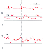 Figure 32-24
Normal central venous pressure (CVP) waveform. The diastolic
components (y descent, end-diastolic a wave) and the systolic components (c wave,
x descent, end-systolic v wave) are all clearly delineated. A mid-diastolic plateau
wave, the h wave, is also seen because the heart rate is slow. Identification of
the waveform is aided by timing the relationship between individual waveform components
and the electrocardiographic R wave. Timing of the waveform with the arterial pressure
(ART) trace is more confusing because of the relative delay in systolic arterial
pressure upstroke. (Redrawn from Mark JB: Atlas of Cardiovascular Monitoring.
New York, Churchill Livingstone, 1998, Fig. 2-5.)
Figure 32-24
Normal central venous pressure (CVP) waveform. The diastolic
components (y descent, end-diastolic a wave) and the systolic components (c wave,
x descent, end-systolic v wave) are all clearly delineated. A mid-diastolic plateau
wave, the h wave, is also seen because the heart rate is slow. Identification of
the waveform is aided by timing the relationship between individual waveform components
and the electrocardiographic R wave. Timing of the waveform with the arterial pressure
(ART) trace is more confusing because of the relative delay in systolic arterial
pressure upstroke. (Redrawn from Mark JB: Atlas of Cardiovascular Monitoring.
New York, Churchill Livingstone, 1998, Fig. 2-5.)
changes in atrial geometry produced by ventricular contraction and ejection. This
is the x descent or systolic collapse in atrial pressure.
The x descent can be divided into two portions, x and x', corresponding to the segments
before and after the c wave. The last atrial pressure peak is the v
wave, which is caused by venous filling of the atrium during late systole
while the tricuspid valve remains closed. The v wave usually peaks just after the
ECG T wave. Atrial pressure then decreases, thereby inscribing the y
descent or diastolic collapse, as the tricuspid valve opens and blood
flows from the atrium to the ventricle. (A final component of the CVP waveform,
the h wave, occasionally appears as a pressure plateau
in mid to late diastole. The h wave is not normally seen unless the heart rate is
slow and venous pressure is elevated.[309]
[310]
)
In summary, the normal venous waveform components may be remembered as follows:
the a wave results from atrial contraction; the c wave from tricuspid valve closure
and isovolumic right ventricular contraction; and the v wave from ventricular ejection,
which drives venous filling of the atrium.
Thus, in relation to the cardiac cycle and ventricular mechanical
actions, the CVP waveform can be considered to have three systolic components (c
wave, x descent, v wave) and two diastolic components (y descent, a wave). By recalling
the mechanical actions that generate the pressure peaks and troughs, it is easy to
identify these waveform components properly by aligning the CVP waveform and the
ECG trace and using the ECG R wave to mark end-diastole and the onset of systole.
When the radial artery pressure trace is used for CVP waveform timing instead of
the ECG, confusion may arise because the arterial pressure upstroke occurs nearly
200 milliseconds after the ECG R wave (see Fig.
32-24
). This normal physiologic delay reflects the times required for
the spread of electrical depolarization through the ventricle (≅60 milliseconds),
isovolumic left ventricular contraction (≅60 milliseconds), transmission of the
increase in aortic pressure to the radial artery (≅50 milliseconds), and transmission
of the increase in radial artery pressure through fluid-filled tubing to the transducer
(≅10 milliseconds).[113]
[136]
The normal CVP peaks are designated systolic (c, v) or diastolic
(a) according to the phase of the cardiac cycle in which the wave begins. However,
one generally identifies these waves not by their onset or upstroke but rather by
the location of their peaks. For instance, the a wave generally begins and peaks
in end-diastole, but the peak may appear delayed to coincide with the ECG R wave,
especially in a patient with a short PR interval. In this instance, the a and c
waves merge, and this composite wave is termed an a-c wave. Designation of the CVP
v wave as a systolic event may be even more confusing. Although ascent of the v
wave begins during late systole, the peak of the v wave occurs during isovolumic
ventricular relaxation, immediately before opening of the atrioventricular valve
and the y descent. Consequently, the most precise description would be that the
v wave begins in late systole but peaks during isovolumic ventricular relaxation,
the earliest portion of diastole. For clinical purposes, it is simplest to consider
the v wave to be a systolic wave.
Although three distinct CVP peaks (a, c, v) and two troughs (x,
y) are discernible in the normal venous pressure trace, heart rate changes and conduction
abnormalities alter this pattern. A short ECG PR interval causes fusion of the a
and c waves, and tachycardia reduces the length of diastole and the duration of the
y descent, which causes the v and a waves to merge. In contrast, bradycardia causes
each wave to become more distinct, with separate x and x' descents visible and a
more prominent h wave. Although in some circumstances other pathologic waves may
be evident in the CVP trace, one should resist the temptation to assign physiologic
significance to each small pressure peak because many will arise as artifacts of
fluid-filled tubing-transducer monitoring systems. Instead, search for the expected
waveform components, including the waveforms that are characteristic of the pathologic
conditions suspected.