|
|
|
|
|
|
|
|
|
|
|
|
|
|
|
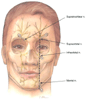 Plate 1
Blockage of the terminal sensory branches of the trigeminal
nerve. A vertical line connects the supraorbital notch, infraorbital foramen, and
mental foramen.
Plate 1
Blockage of the terminal sensory branches of the trigeminal
nerve. A vertical line connects the supraorbital notch, infraorbital foramen, and
mental foramen.
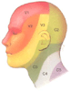 Plate 2
Dermatomes of the head, neck, and face.
Plate 2
Dermatomes of the head, neck, and face.
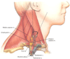 Plate 3
Supraclavicular block. The three trunks are compactly
arranged at the level of the first rib. The needle is systematically walked anteriorly
and posteriorly along the rib until the plexus is located.
Plate 3
Supraclavicular block. The three trunks are compactly
arranged at the level of the first rib. The needle is systematically walked anteriorly
and posteriorly along the rib until the plexus is located.
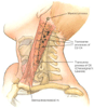 Plate 4
Anatomic landmarks and method of needle placement for
deep cervical plexus blocks at C2, C3, and C4.
Plate 4
Anatomic landmarks and method of needle placement for
deep cervical plexus blocks at C2, C3, and C4.
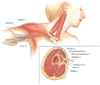 Plate 5
Axillary block. The arm is abducted 90 degrees. Distal
pressure is maintained during needle placement and injection of the local anesthetic.
Plate 5
Axillary block. The arm is abducted 90 degrees. Distal
pressure is maintained during needle placement and injection of the local anesthetic.
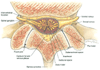 Plate 6
Cross-sectional lumbar vertebral centroneuraxis anatomy
at the L3–4 level.
Plate 6
Cross-sectional lumbar vertebral centroneuraxis anatomy
at the L3–4 level.
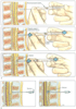 Plate 7
Techniques of epidural needle insertion most often used
in locating the epidural space. A, In the loss-of-resistance
technique, the needle is inserted into the ligamentum flavum, and a syringe containing
an air bubble is attached to the hub. After compression of the air bubble is obtained
by applying pressure to the syringe plunger, the needle is carefully advanced until
its entry into the epidural space is confirmed by the characteristic loss of resistance
to syringe plunger pressure, and the fluid enters the space easily. B,
In the hanging-drop technique, the needle is inserted into the ligamentum flavum,
and a drop of saline (or local anesthetic) is placed in the hub. The needle is then
carefully advanced until its entry into the epidural space is detected by the drop
of solution being "sucked" into the epidural space.
Plate 7
Techniques of epidural needle insertion most often used
in locating the epidural space. A, In the loss-of-resistance
technique, the needle is inserted into the ligamentum flavum, and a syringe containing
an air bubble is attached to the hub. After compression of the air bubble is obtained
by applying pressure to the syringe plunger, the needle is carefully advanced until
its entry into the epidural space is confirmed by the characteristic loss of resistance
to syringe plunger pressure, and the fluid enters the space easily. B,
In the hanging-drop technique, the needle is inserted into the ligamentum flavum,
and a drop of saline (or local anesthetic) is placed in the hub. The needle is then
carefully advanced until its entry into the epidural space is detected by the drop
of solution being "sucked" into the epidural space.
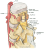 Plate 8
Posterior lumbar vertebral centroneuraxis anatomy.
Plate 8
Posterior lumbar vertebral centroneuraxis anatomy.
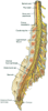 Plate 9
Lateral lumbar vertebral centroneuraxis anatomy from
L1 through the coccyx.
Plate 9
Lateral lumbar vertebral centroneuraxis anatomy from
L1 through the coccyx.
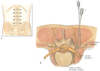 Plate 10
Paravertebral nerve block. A,
Patient position and surface landmarks. B, The needle
is advanced perpendicularly until it contacts the transverse process. It is redirected
to walk off the caudad edge of the transverse process and advanced 1 to 2 cm.
Plate 10
Paravertebral nerve block. A,
Patient position and surface landmarks. B, The needle
is advanced perpendicularly until it contacts the transverse process. It is redirected
to walk off the caudad edge of the transverse process and advanced 1 to 2 cm.
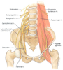 Plate 11
The lumbar plexus lies in the psoas compartment between
the psoas major and quadratus lumborum muscles.
Plate 11
The lumbar plexus lies in the psoas compartment between
the psoas major and quadratus lumborum muscles.
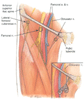 Plate 12
Anatomic landmarks for lateral femoral cutaneous, femoral,
and obturator nerve blocks. In an obturator nerve block, the needle is walked off
the inferior public ramus in a medial and cephalad direction until it passes into
the obturator canal.
Plate 12
Anatomic landmarks for lateral femoral cutaneous, femoral,
and obturator nerve blocks. In an obturator nerve block, the needle is walked off
the inferior public ramus in a medial and cephalad direction until it passes into
the obturator canal.
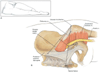 Plate 13
A, Patient positioning.
B, Anatomic landmarks for the posterior approach
to sciatic nerve block.
Plate 13
A, Patient positioning.
B, Anatomic landmarks for the posterior approach
to sciatic nerve block.
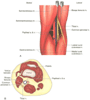 Plate 14
Popliteal fossa block. A,
Anatomic landmarks for the posterior approach to the sciatic nerve in the popliteal
fossa. B, Anatomic landmarks for the lateral approach
to the sciatic nerve in the popliteal fossa.
Plate 14
Popliteal fossa block. A,
Anatomic landmarks for the posterior approach to the sciatic nerve in the popliteal
fossa. B, Anatomic landmarks for the lateral approach
to the sciatic nerve in the popliteal fossa.
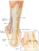 Plate 15
A, Anatomic landmarks
for block of the posterior tibial and sural nerves at the ankle. B,
Posterior tibial nerve and method of needle placement for block at the ankle. C,
Sural nerve and method of needle placement for block at the ankle.
Plate 15
A, Anatomic landmarks
for block of the posterior tibial and sural nerves at the ankle. B,
Posterior tibial nerve and method of needle placement for block at the ankle. C,
Sural nerve and method of needle placement for block at the ankle.
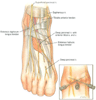 Plate 16
Anatomic landmarks for block of the deep peroneal, superficial
peroneal, and saphenous nerves at the ankle.
Plate 16
Anatomic landmarks for block of the deep peroneal, superficial
peroneal, and saphenous nerves at the ankle.
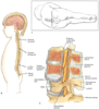 Plate 17
Intervertebral epidural anesthesia. A,
Recommended position of the patient. B, Influence
of spinous processes on needle orientation. C, Anatomy
of the epidural space.
Plate 17
Intervertebral epidural anesthesia. A,
Recommended position of the patient. B, Influence
of spinous processes on needle orientation. C, Anatomy
of the epidural space.
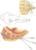 Plate 18
Caudal anesthesia. A,
Anatomic landmarks. B, Position of the patient and
surface landmarks. C, Puncture technique: skin penetration
using a 60- to 90-degree angle to the skin (1), redirection of the needle (2), and
slight penetration (1 to 2 mm) within the spinal canal (3).
Plate 18
Caudal anesthesia. A,
Anatomic landmarks. B, Position of the patient and
surface landmarks. C, Puncture technique: skin penetration
using a 60- to 90-degree angle to the skin (1), redirection of the needle (2), and
slight penetration (1 to 2 mm) within the spinal canal (3).
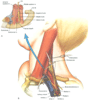 Plate 19
Supraclavicular brachial plexus block. A,
Relationship of the brachial plexus trunks to the clavicle and lower insertions of
the anterior and middle scalene muscles. B, Insertion
routes of the most common supraclavicular approaches to the brachial plexus.
Plate 19
Supraclavicular brachial plexus block. A,
Relationship of the brachial plexus trunks to the clavicle and lower insertions of
the anterior and middle scalene muscles. B, Insertion
routes of the most common supraclavicular approaches to the brachial plexus.
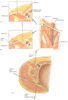 Plate 20
Proximal lower extremity blocks. A,
Fascia iliaca compartment block. B, Classic femoral
nerve block. C, Relationship of the femoral nerve
in the groin. D, Insertion routes of the most common
proximal blocks of the lower extremity (cross section of the thigh).
Plate 20
Proximal lower extremity blocks. A,
Fascia iliaca compartment block. B, Classic femoral
nerve block. C, Relationship of the femoral nerve
in the groin. D, Insertion routes of the most common
proximal blocks of the lower extremity (cross section of the thigh).
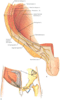 Plate 21
Iliohypogastric and ilioinguinal nerve blocks. A,
Relationship of the ilioinguinal and iliohypogastric nerves. B,
Puncture technique, showing two sites of injection.
Plate 21
Iliohypogastric and ilioinguinal nerve blocks. A,
Relationship of the ilioinguinal and iliohypogastric nerves. B,
Puncture technique, showing two sites of injection.
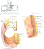 Plate 22
Intercostal nerve blocks. A,
Recommended position of the patient. B, Intercostal
space and puncture technique: insertion of the needle until it contacts the lower
border of the upper rib (1) and caudad redirection of the needle to pass it immediately
below the rib while continuous pressure is exerted on the barrel of the syringe (2).
C, Intercostal nerves and branches.
Plate 22
Intercostal nerve blocks. A,
Recommended position of the patient. B, Intercostal
space and puncture technique: insertion of the needle until it contacts the lower
border of the upper rib (1) and caudad redirection of the needle to pass it immediately
below the rib while continuous pressure is exerted on the barrel of the syringe (2).
C, Intercostal nerves and branches.
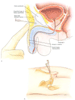 Plate 23
Penile block. A, Relationship
of dorsal nerves and orientation of the needle (sagittal section at the level of
the pubic symphysis). B, Position of the patient
and needle orientation (almost perpendicular to the skin, with a slight slope medially
and caudally).
Plate 23
Penile block. A, Relationship
of dorsal nerves and orientation of the needle (sagittal section at the level of
the pubic symphysis). B, Position of the patient
and needle orientation (almost perpendicular to the skin, with a slight slope medially
and caudally).
|
|
|
|
|
|
|
|
|
|
|
|
|