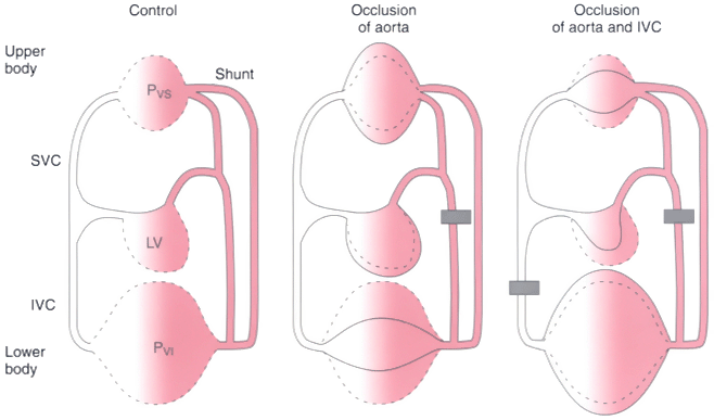
Figure 52-12
Schematic drawing of the circulation. Compliant regions
(stippled lines) of the upper and lower parts of
the body and end-diastolic volumes of the left ventricle in a control state (left
panel) are shown after occlusion of aorta alone (middle
panel) and combined occlusion of the aorta and inferior vena cava (right
panel). IVC, inferior vena cava; LV, left ventricle; PVS
and PVI, pressure in compliant regions of the upper
and lower body respectively; Shunt, physiologic shunt; SVC, superior vena cava.
(From Stokland O, Miller MM, Ilebekk A, Kiil F: Mechanism of hemodynamic
responses to occlusion of the descending thoracic aorta. Am J Physiol 238:H423–H429,
1980.)